Phone: (+33) 625 16 20 71 - Sunday to Friday contact@alliancemedicalfrance.fr
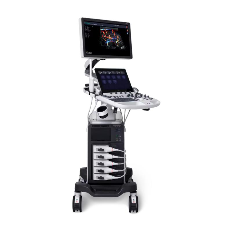
Sonoscape Échographe P40 Elite

Sonoscape

Échographe P40 Elite

Starting from
32 620 .00 €

SonoScape P40 Elite color Doppler ultrasound system with advanced μ-Scan+, SR Flow and 3D/4D imaging, designed to deliver clear, detailed images across diverse clinical applications.

SonoScape P40 Elite – Color Doppler Ultrasound System

Configuration: VC2-9 4D convex, 12LA linear, 6V3 endocavitary.

The SonoScape P40 Elite features a computing architecture combining CPU, GPU, and FPGA to optimize the rendering of fine details and image dynamics. Its optimized analog-to-digital conversion contributes to clearer visualization of subtle anatomical structures.

Probe and Imaging Technologies

  • Single Crystal Probes: provide homogeneous and stable energy transmission.
  • Composite Crystal Linear Probes: support reduced acoustic impedance and improved signal consistency.
  • μ-Scan+: available in B-mode and 3D/4D imaging, improving the distinction between tissue structures and artifacts.
  • SR-Flow: optimizes the separation of blood-flow and tissue signals through dedicated filtering and facilitates the detection of slow flows.
  • Micro F: expands the range of visualizable flows and supports microvascular hemodynamic analysis.

Flow Analysis and Diagnostic Assistance

The system allows flow interpretation to be assessed in relation to surrounding tissues, supporting more detailed evaluation of lesions and tumors.

Several diagnostic assistance and automated quantification tools are integrated:

  • S-Fetus: automated acquisition of standard planes and assistance with fetal biometry.
  • Auto OB: assistance with obstetric measurements.
  • Auto NT: automated nuchal translucency measurement.
  • Auto Bladder: automated bladder volume quantification.
  • Auto IMT: automated intima-media thickness measurement.
  • AVC Follicle: automated follicle quantification.

Optimization and Advanced Applications

Auto B/C and Auto PW functions facilitate the optimization of imaging and pulsed-wave Doppler parameters.

For contrast-enhanced ultrasound, MFI, TIC, and MFI Time provide parametric perfusion analysis. The P40 Elite also incorporates several tools for specialized applications:

  • S-Live and S-Live Silhouette for advanced 3D/4D imaging.
  • Color 3D for volumetric visualization with color mapping.
  • Stress Echo for stress echocardiography examinations.
  • MQA for cardiac analysis and quantification.
Quick delivery worldwide
All our equipment is rigorously tested to ensure its quality and compliance with safety standards.
Download brochure

SonoScape P40 Elite – Color Doppler Ultrasound System

Configuration: VC2-9 4D convex, 12LA linear, 6V3 endocavitary.

The SonoScape P40 Elite features a computing architecture combining CPU, GPU, and FPGA to optimize the rendering of fine details and image dynamics. Its optimized analog-to-digital conversion contributes to clearer visualization of subtle anatomical structures.

Probe and Imaging Technologies

  • Single Crystal Probes: provide homogeneous and stable energy transmission.
  • Composite Crystal Linear Probes: support reduced acoustic impedance and improved signal consistency.
  • μ-Scan+: available in B-mode and 3D/4D imaging, improving the distinction between tissue structures and artifacts.
  • SR-Flow: optimizes the separation of blood-flow and tissue signals through dedicated filtering and facilitates the detection of slow flows.
  • Micro F: expands the range of visualizable flows and supports microvascular hemodynamic analysis.

Flow Analysis and Diagnostic Assistance

The system allows flow interpretation to be assessed in relation to surrounding tissues, supporting more detailed evaluation of lesions and tumors.

Several diagnostic assistance and automated quantification tools are integrated:

  • S-Fetus: automated acquisition of standard planes and assistance with fetal biometry.
  • Auto OB: assistance with obstetric measurements.
  • Auto NT: automated nuchal translucency measurement.
  • Auto Bladder: automated bladder volume quantification.
  • Auto IMT: automated intima-media thickness measurement.
  • AVC Follicle: automated follicle quantification.

Optimization and Advanced Applications

Auto B/C and Auto PW functions facilitate the optimization of imaging and pulsed-wave Doppler parameters.

For contrast-enhanced ultrasound, MFI, TIC, and MFI Time provide parametric perfusion analysis. The P40 Elite also incorporates several tools for specialized applications:

  • S-Live and S-Live Silhouette for advanced 3D/4D imaging.
  • Color 3D for volumetric visualization with color mapping.
  • Stress Echo for stress echocardiography examinations.
  • MQA for cardiac analysis and quantification.
  • μ-Scan+: Enhances contrast and reduces artifacts in 2D and 3D/4D imaging.
  • SR-Flow: Provides sensitive visualization of low-velocity blood flow.
  • Micro F: Facilitates the analysis of microvascular flow for more detailed visualization.
  • S-Fetus: Automates the acquisition of obstetric planes and measurements to simplify examinations.
  • MFI Time: Provides color mapping of different perfusion phases during contrast-enhanced examinations.
  • Sono-synch: Enables real-time sharing of the interface and camera feed to facilitate remote consultations.
Écran :
moniteur 21,5" haute résolution
Cadence système :
FPGA 2× par rapport au système P40 précédent
Réactivité :
GPU 10× par rapport au système P40 précédent
Traitement des données :
CPU 4× par rapport au système P40 précédent
Écran tactile :
13,3"
Autonomie :
batterie pour 2 h de scan continu
  • 3C-A – Convex probe
  • C1-6 – Single-crystal convex probe
  • C613 – Micro-convex probe
  • 12L-A – Linear probe
  • 12L-B – Linear probe
  • 9L-A – Linear probe
Scroll