Phone: (+33) 625 16 20 71 - Sunday to Friday
contact@alliancemedicalfrance.fr
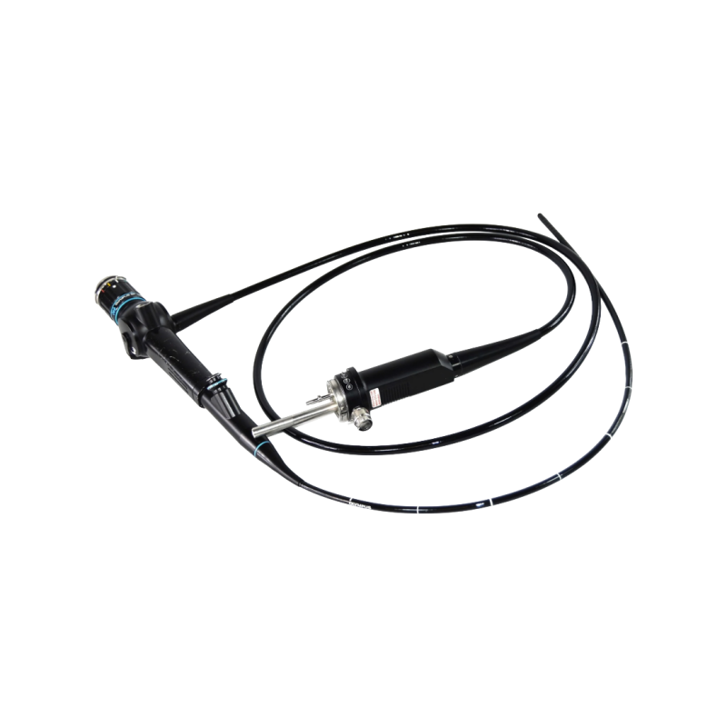
Olympus BF-P30 flexible bronchofibroscope with a 4.9 mm distal diameter, 2.2 mm channel, 550 mm working length and 120° field of view for airway examination.
This device is designed for the endoscopic examination of the airways, with a forward-viewing optical system for examination of the trachea and bronchi. Its optical system provides a 120° field of view and a 3–50 mm depth of field, supporting detailed observation of bronchial structures.
The image can be viewed directly through the eyepiece or transmitted to a compatible endoscopic imaging system. This configuration facilitates anatomical observation as well as documentation of clinical procedures.
The ergonomic control section incorporates an angulation lever that allows the distal tip to be directed during the examination. The bending section responds to the lever movements, facilitating advancement and positioning of the instrument within the airways.
The eyepiece also features a diopter adjustment ring that allows the focus to be adjusted according to the user's requirements. This configuration supports precise control during insertion and endoscopic observation.
The device incorporates an internal instrument channel for the introduction of compatible endoscopic accessories, as well as suction or fluid injection.
A dedicated suction valve allows liquids and debris that may interfere with image visibility to be removed during the examination. A biopsy valve allows compatible instruments or syringes to be introduced for the administration of solutions.
With its optical system, angulation mechanism, and instrument channel, this device supports various diagnostic and therapeutic procedures during bronchoscopic examinations while facilitating visualization and controlled handling of the instrument.
This device is designed for the endoscopic examination of the airways, with a forward-viewing optical system for examination of the trachea and bronchi. Its optical system provides a 120° field of view and a 3–50 mm depth of field, supporting detailed observation of bronchial structures.
The image can be viewed directly through the eyepiece or transmitted to a compatible endoscopic imaging system. This configuration facilitates anatomical observation as well as documentation of clinical procedures.
The ergonomic control section incorporates an angulation lever that allows the distal tip to be directed during the examination. The bending section responds to the lever movements, facilitating advancement and positioning of the instrument within the airways.
The eyepiece also features a diopter adjustment ring that allows the focus to be adjusted according to the user's requirements. This configuration supports precise control during insertion and endoscopic observation.
The device incorporates an internal instrument channel for the introduction of compatible endoscopic accessories, as well as suction or fluid injection.
A dedicated suction valve allows liquids and debris that may interfere with image visibility to be removed during the examination. A biopsy valve allows compatible instruments or syringes to be introduced for the administration of solutions.
With its optical system, angulation mechanism, and instrument channel, this device supports various diagnostic and therapeutic procedures during bronchoscopic examinations while facilitating visualization and controlled handling of the instrument.
 Francais