Phone: (+33) 625 16 20 71 - Sunday to Friday
contact@alliancemedicalfrance.fr
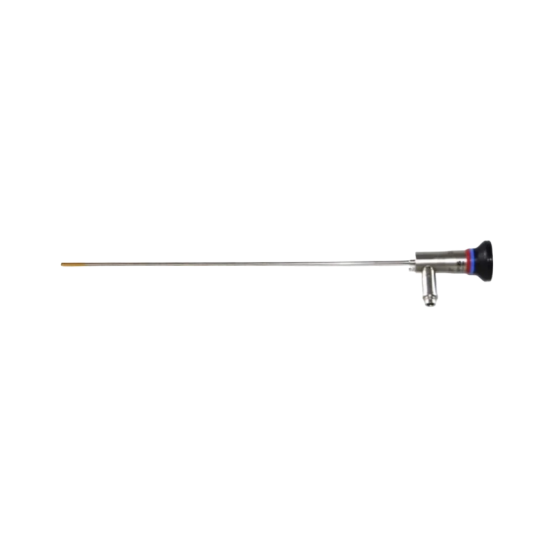
Olympus WA20017A rigid telescope with 4 mm diameter, 30° wide-angle view and autoclavable design. It uses rod-lens optics and fiber-optic illumination for urological endoscopic visualization.
The WA20017A is designed for urological endoscopic procedures requiring precise visualization of urinary structures. It allows visual examination during diagnostic procedures as well as operative interventions.
Its optical system is based on a rod-lens architecture that transmits the image from the examined area to the eyepiece or a compatible video device. Illumination of the operative area is provided by an optical fiber bundle connected to an external light source.
The telescope is inserted into the dedicated telescope channel of a compatible endoscopic instrument and secured using a dedicated connector. The assembly includes an insertion tube, light-guide connector, eyepiece, and color-coded markings to facilitate instrument identification in the operating environment.
Connection to a video adapter or camera head allows the image to be transmitted to a visualization system used during diagnostic and interventional procedures.
Before each use, the instrument should be inspected to verify image quality and proper light transmission. The visual inspection is performed at a 15 mm test distance to check image sharpness and confirm that there is no apparent optical deterioration.
The WA20017A is a precision optical instrument that requires careful handling and inspection to help preserve its visual performance during endoscopic procedures.
The WA20017A is designed for urological endoscopic procedures requiring precise visualization of urinary structures. It allows visual examination during diagnostic procedures as well as operative interventions.
Its optical system is based on a rod-lens architecture that transmits the image from the examined area to the eyepiece or a compatible video device. Illumination of the operative area is provided by an optical fiber bundle connected to an external light source.
The telescope is inserted into the dedicated telescope channel of a compatible endoscopic instrument and secured using a dedicated connector. The assembly includes an insertion tube, light-guide connector, eyepiece, and color-coded markings to facilitate instrument identification in the operating environment.
Connection to a video adapter or camera head allows the image to be transmitted to a visualization system used during diagnostic and interventional procedures.
Before each use, the instrument should be inspected to verify image quality and proper light transmission. The visual inspection is performed at a 15 mm test distance to check image sharpness and confirm that there is no apparent optical deterioration.
The WA20017A is a precision optical instrument that requires careful handling and inspection to help preserve its visual performance during endoscopic procedures.
 Francais