Phone: (+33) 625 16 20 71 - Sunday to Friday
contact@alliancemedicalfrance.fr
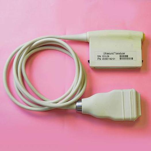
The Philips L12-3 is a broadband linear array probe (12–3 MHz, 160 elements, 38 mm aperture) for vascular, MSK, small parts and superficial imaging. The Ergo version adds an ergonomic grip, MicroFlow Imaging and XRES Pro. Biopsy capable.
The Philips L12-3 is a broadband linear-array ultrasound transducer designed for detailed imaging of superficial and relatively shallow anatomical structures. Its 12–3 MHz frequency range provides a balance between high-resolution imaging and penetration, making it suitable for a broad range of vascular, musculoskeletal, superficial, and interventional examinations.
The L12-3 supports several imaging and Doppler modes designed to provide detailed anatomical visualization and assessment of blood flow. Philips documents the following modes and imaging technologies for this transducer:
The Philips L12-3 is designed for a wide range of examinations involving superficial and moderately deep structures. Its linear configuration is particularly suitable when high-resolution visualization is required.
The L12-3 is particularly suited to vascular ultrasound. Its linear-array geometry and broadband frequency range support detailed visualization of carotid, arterial, and venous structures. Color and pulsed-wave Doppler capabilities can be used for assessment of blood flow when supported by the ultrasound system.
The high-frequency capability of the L12-3 makes it suitable for imaging superficial anatomical structures. It can be used for musculoskeletal examinations and small-parts imaging where detailed visualization of superficial tissues is required.
The L12-3 is documented for interventional applications and is biopsy capable. Its linear design can provide a practical imaging field for procedures requiring ultrasound guidance.
The broadband design provides an operating range from 3 to 12 MHz. Lower frequencies within the range can support greater penetration, while higher frequencies can provide increased detail for superficial structures. This makes the transducer adaptable to different examination requirements.
Philips specifically identifies the L12-3 as biopsy capable. This allows the transducer to be used with appropriate compatible biopsy accessories for ultrasound-guided procedures.
The Philips specifications identify the L12-3 as not capable of Image Fusion Navigation. It should therefore not be described as supporting image-fusion navigation without additional system-specific documentation.
Philips does not provide a numerical field-of-view value for the L12-3 in the official specification currently available. Therefore, no numerical field-of-view measurement is included here.
The Philips L12-3 is a broadband linear-array ultrasound transducer designed for detailed imaging of superficial and relatively shallow anatomical structures. Its 12–3 MHz frequency range provides a balance between high-resolution imaging and penetration, making it suitable for a broad range of vascular, musculoskeletal, superficial, and interventional examinations.
The L12-3 supports several imaging and Doppler modes designed to provide detailed anatomical visualization and assessment of blood flow. Philips documents the following modes and imaging technologies for this transducer:
The Philips L12-3 is designed for a wide range of examinations involving superficial and moderately deep structures. Its linear configuration is particularly suitable when high-resolution visualization is required.
The L12-3 is particularly suited to vascular ultrasound. Its linear-array geometry and broadband frequency range support detailed visualization of carotid, arterial, and venous structures. Color and pulsed-wave Doppler capabilities can be used for assessment of blood flow when supported by the ultrasound system.
The high-frequency capability of the L12-3 makes it suitable for imaging superficial anatomical structures. It can be used for musculoskeletal examinations and small-parts imaging where detailed visualization of superficial tissues is required.
The L12-3 is documented for interventional applications and is biopsy capable. Its linear design can provide a practical imaging field for procedures requiring ultrasound guidance.
The broadband design provides an operating range from 3 to 12 MHz. Lower frequencies within the range can support greater penetration, while higher frequencies can provide increased detail for superficial structures. This makes the transducer adaptable to different examination requirements.
Philips specifically identifies the L12-3 as biopsy capable. This allows the transducer to be used with appropriate compatible biopsy accessories for ultrasound-guided procedures.
The Philips specifications identify the L12-3 as not capable of Image Fusion Navigation. It should therefore not be described as supporting image-fusion navigation without additional system-specific documentation.
Philips does not provide a numerical field-of-view value for the L12-3 in the official specification currently available. Therefore, no numerical field-of-view measurement is included here.
 Francais