Phone: (+33) 625 16 20 71 - Sunday to Friday
contact@alliancemedicalfrance.fr
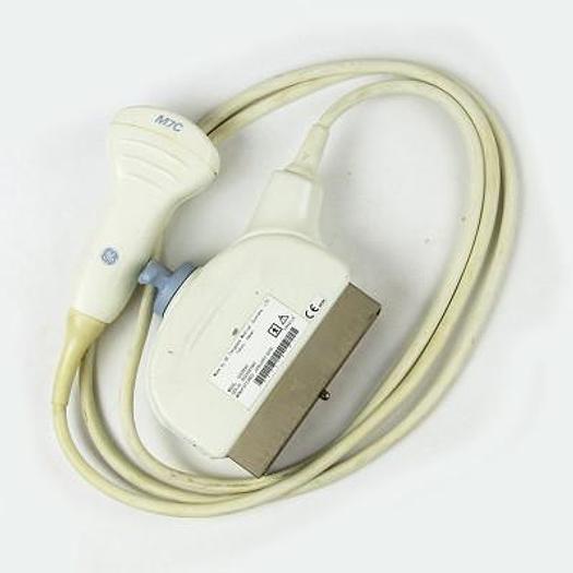
GE HealthCare M7C Matrix convex probe for abdominal, OB/GYN and pediatric imaging. It offers a 2.5–8.0 MHz bandwidth and 62° field of view for versatile examinations.
The GE M7C is a matrix-array convex ultrasound transducer designed for general-purpose diagnostic imaging. It is intended primarily for abdominal, obstetric, gynecological, and pediatric examinations. Its wide frequency bandwidth and convex scanning geometry make it suitable for imaging both relatively deep anatomical structures and routine general-imaging applications.
The M7C uses a matrix-array design rather than a conventional single-row curved array. GE documentation identifies 960 elements and a compact 58 × 18 mm footprint. This configuration is designed to provide flexible imaging performance across a range of general diagnostic applications.
The convex geometry provides a broad imaging window while the relatively low-frequency operating range supports penetration into deeper anatomical regions. This makes the M7C particularly suitable for abdominal imaging and other examinations where depth is important.
The M7C is documented with a frequency bandwidth of approximately 2.5 to 8.0 MHz. GE technical documentation specifies a maximum bandwidth of 2.6–7.7 MHz at -20 dB. The available bandwidth allows the system to select appropriate operating frequencies according to the examination and imaging depth.
The combination of a broad bandwidth, convex footprint, and matrix-array construction makes the probe suitable for both routine general imaging and examinations requiring deeper penetration.
The GE M7C is primarily intended for abdominal ultrasound. Its convex configuration and frequency range are well suited to imaging deeper abdominal structures and organs.
GE documentation lists the M7C for obstetric and gynecological applications. Its convex scanning geometry provides a broad imaging field that can be useful for routine fetal and pelvic examinations when used with a compatible ultrasound system.
The M7C is also documented for pediatric applications. Its bandwidth and convex design allow it to be used for general pediatric imaging where a suitable convex transducer is required.
On compatible GE LOGIQ systems, the M7C can be used with a range of system-dependent imaging functions. GE LOGIQ 7 documentation lists support for features including Coded Excitation, Coded Harmonics, B-Flow, Coded Contrast, LOGIQ View, Virtual Convex, Easy 3D, Advanced 3D, Anatomical M-Mode, M Color Flow, TruAccess, and biopsy functionality with the M7C. Availability of individual functions depends on the ultrasound system and software configuration.
GE documentation identifies the M7C with a multi-angle reusable biopsy guide. This configuration is intended to support needle-guided procedures when the appropriate biopsy accessory and compatible system configuration are used.
With its compact 58 × 18 mm footprint and convex scanning surface, the M7C is designed to provide a practical balance between patient contact area, imaging coverage, and penetration. Its matrix-array construction and wide bandwidth make it a versatile transducer for general diagnostic ultrasound.
The GE M7C is a matrix-array convex ultrasound transducer designed for general-purpose diagnostic imaging. It is intended primarily for abdominal, obstetric, gynecological, and pediatric examinations. Its wide frequency bandwidth and convex scanning geometry make it suitable for imaging both relatively deep anatomical structures and routine general-imaging applications.
The M7C uses a matrix-array design rather than a conventional single-row curved array. GE documentation identifies 960 elements and a compact 58 × 18 mm footprint. This configuration is designed to provide flexible imaging performance across a range of general diagnostic applications.
The convex geometry provides a broad imaging window while the relatively low-frequency operating range supports penetration into deeper anatomical regions. This makes the M7C particularly suitable for abdominal imaging and other examinations where depth is important.
The M7C is documented with a frequency bandwidth of approximately 2.5 to 8.0 MHz. GE technical documentation specifies a maximum bandwidth of 2.6–7.7 MHz at -20 dB. The available bandwidth allows the system to select appropriate operating frequencies according to the examination and imaging depth.
The combination of a broad bandwidth, convex footprint, and matrix-array construction makes the probe suitable for both routine general imaging and examinations requiring deeper penetration.
The GE M7C is primarily intended for abdominal ultrasound. Its convex configuration and frequency range are well suited to imaging deeper abdominal structures and organs.
GE documentation lists the M7C for obstetric and gynecological applications. Its convex scanning geometry provides a broad imaging field that can be useful for routine fetal and pelvic examinations when used with a compatible ultrasound system.
The M7C is also documented for pediatric applications. Its bandwidth and convex design allow it to be used for general pediatric imaging where a suitable convex transducer is required.
On compatible GE LOGIQ systems, the M7C can be used with a range of system-dependent imaging functions. GE LOGIQ 7 documentation lists support for features including Coded Excitation, Coded Harmonics, B-Flow, Coded Contrast, LOGIQ View, Virtual Convex, Easy 3D, Advanced 3D, Anatomical M-Mode, M Color Flow, TruAccess, and biopsy functionality with the M7C. Availability of individual functions depends on the ultrasound system and software configuration.
GE documentation identifies the M7C with a multi-angle reusable biopsy guide. This configuration is intended to support needle-guided procedures when the appropriate biopsy accessory and compatible system configuration are used.
With its compact 58 × 18 mm footprint and convex scanning surface, the M7C is designed to provide a practical balance between patient contact area, imaging coverage, and penetration. Its matrix-array construction and wide bandwidth make it a versatile transducer for general diagnostic ultrasound.
 Francais