Phone: (+33) 625 16 20 71 - Sunday to Friday
contact@alliancemedicalfrance.fr
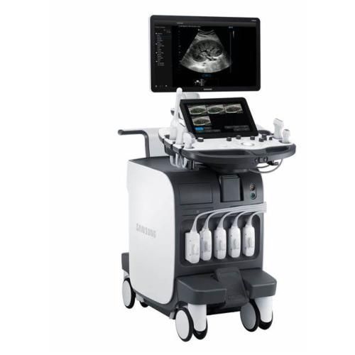
The RS80A is a cart-based ultrasound system from Samsung Medison, using S-Vision/S-Vue imaging, Clear Track needle guidance, QuickScan Doppler optimization, and Realistic Vue 3D rendering, with a broad transducer lineup.
The RS80A is a full-size, cart-based ultrasound system from Samsung Medison, aimed at general imaging, vascular, musculoskeletal, and cardiac use. It's built around two core technologies: a signal-processing architecture called S-Vision, and a matching line of transducers called S-Vue. The rest of the system — control panel, monitor, transducer bay — is designed around making those two things easier to use during a scan.
S-Vision covers two separate stages of image formation. The beamformer handles signal transmission and reception first. Samsung's marketing materials show a side-by-side comparison against a "conventional" beamformer, with the RS80A output described as tighter and less scattered — though the conventional example isn't specified, so that particular claim is worth treating with some skepticism.
The second stage, the imaging engine, processes the beamformed signal to reduce noise while trying to preserve resolution and tissue uniformity. The brochure frames this as raising resolution, penetration, and uniformity while pushing noise down — standard goals for any image processing pipeline, described here without much detail on how they're achieved.
S-Vue transducers use a wider bandwidth design. The brochure explains this as combining the frequency responses of what would otherwise be two separate transducers into one broader band, meant to improve penetration and sensitivity together rather than trading one for the other. The CA1-7A curved array is shown as an example, used for an abdominal scan.
Clear Track is Samsung's needle-visualization feature for interventional procedures — biopsies, injections, and similar guided work. A needle sensor projects the needle's path on screen in real time, regardless of the angle it enters at.
Advanced QuickScan is a one-button optimization for gray scale and Doppler settings — color gain, color box position, and related parameters adjust automatically when pressed. The brochure's example is a carotid artery Doppler scan, comparing images before and after QuickScan; the "after" image shows a noticeably tighter, more centered color box.
Realistic Vue is the system's 3D rendering mode. It renders 3D anatomy with a user-adjustable light source, which creates shadowing to help distinguish surface structures — useful for OB/GYN work in particular, where surface anatomy benefits from depth cues that flat grayscale doesn't provide.
The system has a 23" full HD LED display, described as holding contrast and color across different room lighting, plus a 13.3" tilting touch screen that adjusts to the operator's position. The main monitor folds down flat for transport between rooms. The control panel adjusts on six axes for height, tilt, and rotation, and returns to a home position automatically on power-down, which makes the cart easier to move. Console buttons are grouped around a 3D joystick for simpler navigation during a scan, and a foot pedal locks or releases the console's swivel.
The brochure's image gallery includes examples across a range of applications — fatty liver, gallbladder stone, wrist vessel, finger ganglion, carotid artery with color Doppler, breast mass, transverse right ovary, thyroid nodule with Doppler, a 4-chamber cardiac view, quadriceps, wrist ganglion, and a panoramic musculoskeletal image. These are presented as representative output rather than a specific patient study.
The RS80A is a full-size, cart-based ultrasound system from Samsung Medison, aimed at general imaging, vascular, musculoskeletal, and cardiac use. It's built around two core technologies: a signal-processing architecture called S-Vision, and a matching line of transducers called S-Vue. The rest of the system — control panel, monitor, transducer bay — is designed around making those two things easier to use during a scan.
S-Vision covers two separate stages of image formation. The beamformer handles signal transmission and reception first. Samsung's marketing materials show a side-by-side comparison against a "conventional" beamformer, with the RS80A output described as tighter and less scattered — though the conventional example isn't specified, so that particular claim is worth treating with some skepticism.
The second stage, the imaging engine, processes the beamformed signal to reduce noise while trying to preserve resolution and tissue uniformity. The brochure frames this as raising resolution, penetration, and uniformity while pushing noise down — standard goals for any image processing pipeline, described here without much detail on how they're achieved.
S-Vue transducers use a wider bandwidth design. The brochure explains this as combining the frequency responses of what would otherwise be two separate transducers into one broader band, meant to improve penetration and sensitivity together rather than trading one for the other. The CA1-7A curved array is shown as an example, used for an abdominal scan.
Clear Track is Samsung's needle-visualization feature for interventional procedures — biopsies, injections, and similar guided work. A needle sensor projects the needle's path on screen in real time, regardless of the angle it enters at.
Advanced QuickScan is a one-button optimization for gray scale and Doppler settings — color gain, color box position, and related parameters adjust automatically when pressed. The brochure's example is a carotid artery Doppler scan, comparing images before and after QuickScan; the "after" image shows a noticeably tighter, more centered color box.
Realistic Vue is the system's 3D rendering mode. It renders 3D anatomy with a user-adjustable light source, which creates shadowing to help distinguish surface structures — useful for OB/GYN work in particular, where surface anatomy benefits from depth cues that flat grayscale doesn't provide.
The system has a 23" full HD LED display, described as holding contrast and color across different room lighting, plus a 13.3" tilting touch screen that adjusts to the operator's position. The main monitor folds down flat for transport between rooms. The control panel adjusts on six axes for height, tilt, and rotation, and returns to a home position automatically on power-down, which makes the cart easier to move. Console buttons are grouped around a 3D joystick for simpler navigation during a scan, and a foot pedal locks or releases the console's swivel.
The brochure's image gallery includes examples across a range of applications — fatty liver, gallbladder stone, wrist vessel, finger ganglion, carotid artery with color Doppler, breast mass, transverse right ovary, thyroid nodule with Doppler, a 4-chamber cardiac view, quadriceps, wrist ganglion, and a panoramic musculoskeletal image. These are presented as representative output rather than a specific patient study.
 Francais