Phone: (+33) 625 16 20 71 - Sunday to Friday contact@alliancemedicalfrance.fr
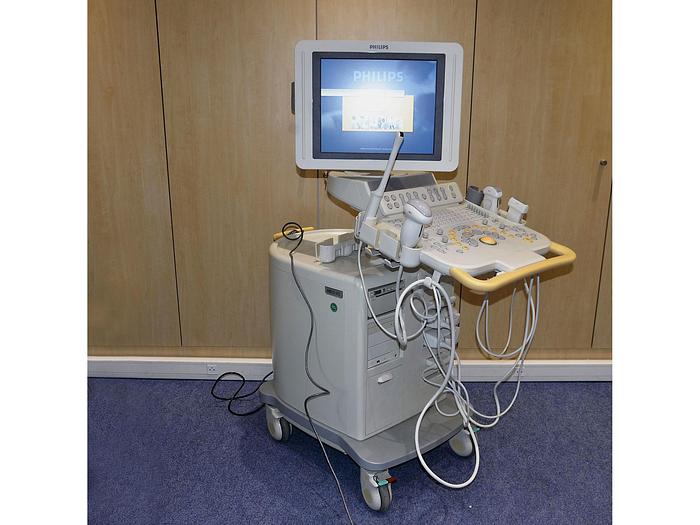
Philips ECHOGRAPHE PHILIPS HD 11 XE

Philips

ECHOGRAPHE PHILIPS HD 11 XE

Starting from
8 000 .00 €

Philips HD11 XE diagnostic ultrasound system designed for versatile clinical imaging, supporting advanced 2D, Doppler and harmonic imaging across a wide range of applications.

Philips HD11 XE – Ultrasound System

The Philips HD11 XE is a complete digital ultrasound system designed to provide high-definition imaging, versatile clinical applications, and an efficient workflow in a compact and reliable platform. Philips developed the HD11 XE for use in hospitals, satellite clinics, specialized practices, and shared-service imaging environments.

Advanced Imaging Technology

  • Broadband beamforming: Provides advanced ultrasound signal processing across a wide range of clinical applications.
  • SonoCT: Uses beam-steered spatial compounding in transmit and receive to acquire multiple lines of sight and combine them in real time, helping produce clearer and more uniform images.
  • XRES: Adaptive image-processing technology designed to reduce artifacts and improve the definition of anatomical borders and margins.
  • Pulse Inversion Harmonic Imaging: Provides broadband harmonic imaging for enhanced grayscale presentation.
  • Adaptive Color Doppler: Automatically selects an appropriate Doppler or angio frequency to support sensitive flow visualization.
  • Color Power Angio: Provides visualization of flow amplitude and direction.
  • Adaptive Doppler: Designed to enhance weak Doppler signals and reduce noise.

Imaging Modes

The HD11 XE provides a broad range of imaging capabilities to support general imaging, vascular, cardiac, and other specialized examinations, depending on the system configuration and installed options.

  • 2D grayscale imaging
  • Pulse Inversion Harmonic Imaging
  • M-mode
  • Anatomical M-mode
  • Color Doppler
  • Color Power Angio
  • Pulsed Wave Doppler
  • Continuous Wave Doppler
  • Tissue Doppler Imaging (TDI)
  • Color TDI
  • High-PRF Doppler

Cardiology and Cardiovascular Imaging

The HD11 XE can be configured for cardiovascular applications and offers technologies designed to support cardiac assessment. Philips documentation describes capabilities including Tissue Doppler Imaging, Color TDI, Anatomical M-mode, and Doppler technologies.

  • Anatomical M-mode: Facilitates measurements of cardiac chambers, walls, and ejection fraction by allowing the M-mode line to be positioned relative to anatomy.
  • Tissue Doppler Imaging: Supports assessment of myocardial motion and tissue velocities.
  • Color TDI: Provides visualization of the direction and timing of myocardial motion.
  • Stress Echo: Available as an integrated option for acquisition and review of stress echocardiography studies.
  • Contrast imaging: Available as an option for selected applications and compatible transducers.

Vascular Imaging

The HD11 XE supports vascular examinations with dedicated Doppler and imaging technologies. Its vascular capabilities can be used for assessment of vessels and abdominal structures, with optional features available for more specialized examinations.

  • Color Doppler and spectral Doppler for blood-flow assessment
  • Adaptive Doppler technology for improved weak-signal detection
  • Trapezoidal imaging for an expanded field of view with compatible linear transducers
  • Intelligent Doppler for angle-consistent spectral Doppler measurements
  • Optional QLAB IMT functionality for intima-media thickness measurements
  • Transcranial Doppler solutions available with compatible transducers

General Imaging and Clinical Applications

The HD11 XE is designed as a versatile platform that can be configured for a broad range of clinical environments. Its application portfolio includes general imaging and specialized examinations depending on the installed software, transducer, and options.

  • Abdominal imaging
  • Small-parts imaging
  • Musculoskeletal examinations
  • Obstetrics and gynecology
  • Pediatric imaging
  • Vascular imaging
  • Cardiology
  • Intraoperative imaging
  • Transcranial Doppler
  • Prostate and urological applications
  • Emergency and specialized clinical applications, depending on configuration

3D and Panoramic Imaging

  • 3D imaging: The HD11 XE supports 3D imaging capabilities with multiplanar visualization and interactive viewing of acquired volume data, depending on configuration.
  • Panoramic imaging: An optional extended-field-of-view function can create a larger panoramic representation by combining images acquired while moving the transducer across the anatomy.

Workflow and Image Optimization

Philips designed the HD11 XE to combine advanced imaging with an efficient clinical workflow. Automated optimization and image-processing technologies help simplify examinations while maintaining access to detailed imaging controls.

  • Automated image optimization: Helps streamline adjustment of imaging parameters.
  • Image thumbnails: Allow users to review acquired images and monitor the progress of an examination.
  • Exam management: Supports recording, reviewing, editing, archiving, and documentation of ultrasound examinations.
  • Report creation: Supports examination reports with embedded images.

Data Management and Connectivity

The HD11 XE provides data-management and documentation capabilities intended to support clinical workflow. Philips documentation also lists DICOM conformance information for multiple HD11 XE software revisions, including R1.1.x.x, R1.2.x, and R2.0.

  • DICOM support depending on software configuration
  • Image recording and archiving
  • Image review and editing
  • Exam documentation
  • Report creation with embedded images
  • CD-based documentation and archiving capabilities
  • Support for optional peripheral devices

Expandable Platform

A key characteristic of the HD11 XE is its expandable architecture. Philips designed the system so that additional clinical capabilities could be added according to the requirements of the facility.

  • Cardiac and stress echocardiography capabilities
  • Advanced quantification with QLAB, depending on configuration
  • Contrast imaging options
  • Transesophageal echocardiography capabilities
  • Vascular imaging options
  • Panoramic imaging
  • 3D imaging capabilities

Clinical Environment

The Philips HD11 XE is suitable for healthcare facilities requiring a versatile ultrasound platform capable of supporting multiple departments and clinical specialties. Philips specifically describes the system as suitable for public and private hospitals, satellite clinics, specialized practices, shared-service laboratories, private offices, and mobile imaging services.

Key Features

  • Philips HD11 XE digital ultrasound platform
  • High-definition ultrasound imaging
  • Broadband beamforming
  • SonoCT spatial compounding
  • XRES adaptive image processing
  • Pulse Inversion Harmonic Imaging
  • Adaptive Color Doppler
  • Color Power Angio
  • Pulsed Wave and Continuous Wave Doppler
  • Adaptive Doppler technology
  • Tissue Doppler Imaging
  • Anatomical M-mode
  • 3D imaging capabilities
  • Optional panoramic imaging
  • Cardiac and stress echocardiography capabilities
  • Flexible data management and reporting
  • DICOM support depending on software configuration
  • Upgradeable platform designed for multiple clinical environments

Product Status

The Philips HD11 XE is a discontinued ultrasound platform, but Philips continues to provide historical documentation and support information for the system. Official Philips documentation identifies several HD11 XE software revisions, including R1.1.x.x, R1.2.x, and R2.0.

Quick delivery worldwide
All our equipment is rigorously tested to ensure its quality and compliance with safety standards.
Download brochure

Philips HD11 XE – Ultrasound System

The Philips HD11 XE is a complete digital ultrasound system designed to provide high-definition imaging, versatile clinical applications, and an efficient workflow in a compact and reliable platform. Philips developed the HD11 XE for use in hospitals, satellite clinics, specialized practices, and shared-service imaging environments.

Advanced Imaging Technology

  • Broadband beamforming: Provides advanced ultrasound signal processing across a wide range of clinical applications.
  • SonoCT: Uses beam-steered spatial compounding in transmit and receive to acquire multiple lines of sight and combine them in real time, helping produce clearer and more uniform images.
  • XRES: Adaptive image-processing technology designed to reduce artifacts and improve the definition of anatomical borders and margins.
  • Pulse Inversion Harmonic Imaging: Provides broadband harmonic imaging for enhanced grayscale presentation.
  • Adaptive Color Doppler: Automatically selects an appropriate Doppler or angio frequency to support sensitive flow visualization.
  • Color Power Angio: Provides visualization of flow amplitude and direction.
  • Adaptive Doppler: Designed to enhance weak Doppler signals and reduce noise.

Imaging Modes

The HD11 XE provides a broad range of imaging capabilities to support general imaging, vascular, cardiac, and other specialized examinations, depending on the system configuration and installed options.

  • 2D grayscale imaging
  • Pulse Inversion Harmonic Imaging
  • M-mode
  • Anatomical M-mode
  • Color Doppler
  • Color Power Angio
  • Pulsed Wave Doppler
  • Continuous Wave Doppler
  • Tissue Doppler Imaging (TDI)
  • Color TDI
  • High-PRF Doppler

Cardiology and Cardiovascular Imaging

The HD11 XE can be configured for cardiovascular applications and offers technologies designed to support cardiac assessment. Philips documentation describes capabilities including Tissue Doppler Imaging, Color TDI, Anatomical M-mode, and Doppler technologies.

  • Anatomical M-mode: Facilitates measurements of cardiac chambers, walls, and ejection fraction by allowing the M-mode line to be positioned relative to anatomy.
  • Tissue Doppler Imaging: Supports assessment of myocardial motion and tissue velocities.
  • Color TDI: Provides visualization of the direction and timing of myocardial motion.
  • Stress Echo: Available as an integrated option for acquisition and review of stress echocardiography studies.
  • Contrast imaging: Available as an option for selected applications and compatible transducers.

Vascular Imaging

The HD11 XE supports vascular examinations with dedicated Doppler and imaging technologies. Its vascular capabilities can be used for assessment of vessels and abdominal structures, with optional features available for more specialized examinations.

  • Color Doppler and spectral Doppler for blood-flow assessment
  • Adaptive Doppler technology for improved weak-signal detection
  • Trapezoidal imaging for an expanded field of view with compatible linear transducers
  • Intelligent Doppler for angle-consistent spectral Doppler measurements
  • Optional QLAB IMT functionality for intima-media thickness measurements
  • Transcranial Doppler solutions available with compatible transducers

General Imaging and Clinical Applications

The HD11 XE is designed as a versatile platform that can be configured for a broad range of clinical environments. Its application portfolio includes general imaging and specialized examinations depending on the installed software, transducer, and options.

  • Abdominal imaging
  • Small-parts imaging
  • Musculoskeletal examinations
  • Obstetrics and gynecology
  • Pediatric imaging
  • Vascular imaging
  • Cardiology
  • Intraoperative imaging
  • Transcranial Doppler
  • Prostate and urological applications
  • Emergency and specialized clinical applications, depending on configuration

3D and Panoramic Imaging

  • 3D imaging: The HD11 XE supports 3D imaging capabilities with multiplanar visualization and interactive viewing of acquired volume data, depending on configuration.
  • Panoramic imaging: An optional extended-field-of-view function can create a larger panoramic representation by combining images acquired while moving the transducer across the anatomy.

Workflow and Image Optimization

Philips designed the HD11 XE to combine advanced imaging with an efficient clinical workflow. Automated optimization and image-processing technologies help simplify examinations while maintaining access to detailed imaging controls.

  • Automated image optimization: Helps streamline adjustment of imaging parameters.
  • Image thumbnails: Allow users to review acquired images and monitor the progress of an examination.
  • Exam management: Supports recording, reviewing, editing, archiving, and documentation of ultrasound examinations.
  • Report creation: Supports examination reports with embedded images.

Data Management and Connectivity

The HD11 XE provides data-management and documentation capabilities intended to support clinical workflow. Philips documentation also lists DICOM conformance information for multiple HD11 XE software revisions, including R1.1.x.x, R1.2.x, and R2.0.

  • DICOM support depending on software configuration
  • Image recording and archiving
  • Image review and editing
  • Exam documentation
  • Report creation with embedded images
  • CD-based documentation and archiving capabilities
  • Support for optional peripheral devices

Expandable Platform

A key characteristic of the HD11 XE is its expandable architecture. Philips designed the system so that additional clinical capabilities could be added according to the requirements of the facility.

  • Cardiac and stress echocardiography capabilities
  • Advanced quantification with QLAB, depending on configuration
  • Contrast imaging options
  • Transesophageal echocardiography capabilities
  • Vascular imaging options
  • Panoramic imaging
  • 3D imaging capabilities

Clinical Environment

The Philips HD11 XE is suitable for healthcare facilities requiring a versatile ultrasound platform capable of supporting multiple departments and clinical specialties. Philips specifically describes the system as suitable for public and private hospitals, satellite clinics, specialized practices, shared-service laboratories, private offices, and mobile imaging services.

Key Features

  • Philips HD11 XE digital ultrasound platform
  • High-definition ultrasound imaging
  • Broadband beamforming
  • SonoCT spatial compounding
  • XRES adaptive image processing
  • Pulse Inversion Harmonic Imaging
  • Adaptive Color Doppler
  • Color Power Angio
  • Pulsed Wave and Continuous Wave Doppler
  • Adaptive Doppler technology
  • Tissue Doppler Imaging
  • Anatomical M-mode
  • 3D imaging capabilities
  • Optional panoramic imaging
  • Cardiac and stress echocardiography capabilities
  • Flexible data management and reporting
  • DICOM support depending on software configuration
  • Upgradeable platform designed for multiple clinical environments

Product Status

The Philips HD11 XE is a discontinued ultrasound platform, but Philips continues to provide historical documentation and support information for the system. Official Philips documentation identifies several HD11 XE software revisions, including R1.1.x.x, R1.2.x, and R2.0.

Modèle :
HD 11 XE
  • C5-2 – Ref. 21426A
  • C6-3 – Ref. 989605359591
  • C8-4v – Ref. 21437A
  • C8-5 – Ref. 8500-1664-01
  • C9-4 – Ref. 8500-1658-01
  • C9-5ec – Ref. 8500-1655-01
  • PA4-2 – Ref. 21422A
Scroll