Phone: (+33) 625 16 20 71 - Sunday to Friday
contact@alliancemedicalfrance.fr
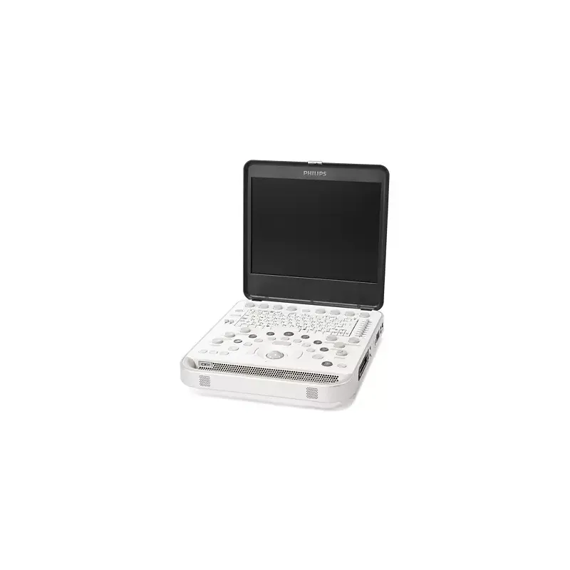
Philips CX50 CompactXtreme: a portable ultrasound system for cardiac, vascular, abdominal, pediatric and OB/GYN exams. Features PureWave transducers, Live 3D imaging, and cart, travel case or hand-carry configurations for use anywhere.
The CX50 CompactXtreme is a compact ultrasound system built on Philips' proprietary imaging platform. It uses PureWave crystal technology, previously limited to Philips' premium systems, to capture a broad band of tissue information. AutoSCAN optimization and tissue-specific presets adjust image settings automatically by exam type, and the system is fully configurable: imaging capabilities, transducers, and analysis tools can be selected to match specific clinical needs, with upgrades available later.
The system supports a wide range of exams, including:
The CX50 offers 2D grayscale, M-mode, anatomical M-mode, Live xPlane, and Live 3D TEE imaging with color flow. Doppler options include pulsed wave, continuous wave, spectral, color, tissue, and Color Power Angio Doppler. Contrast imaging is supported on select transducers, with pulse inversion, power modulation, and coded harmonic modes. Tissue Harmonic Imaging, SonoCT compound imaging, and XRES adaptive processing are available across most transducers to reduce artifacts and improve clarity.
Available on the X7-2t transducer, this mode provides live full-volume imaging, 3D color flow, beat-by-beat retrospective volume selection, and basic on-image measurements, along with ECG display and dynamic face cropping.
Qualitative 3D volume acquisition is supported on all imaging transducers, with surface rendering, multiplanar view display, and QLAB volume analysis tools such as 3DQa and iSlice.
The CX50 supports curved, linear, and sector array transducers, plus a dedicated connector for continuous wave Doppler (Pedoff) transducers. PureWave technology is available in curved, sector, and xMATRIX configurations up to 12 MHz. Transducer options include:
An IntraCardiac Echocardiography (ICE) option operates with St. Jude Medical's patient interface module and Viewflex Xtra catheter for structural heart disease and EP procedures.
The system includes standard measurement tools plus QLAB advanced quantification plug-ins, covering 3D cardiac quantification, cardiac motion and mechanics, mitral valve quantification, strain quantification, intima media thickness measurement, and microvascular and general imaging 3D quantification.
On-screen annotation, SmartExam guided protocols, cineloop review, and stress echo protocols support routine and specialized workflows. Connectivity includes DICOM print, store and structured reporting, DHCP networking, wireless "B and G" networking, and two USB ports. The system can be configured for portability with a specially designed mobility cart, a wheeled travel case, or hand-carry use with an additional AC adapter, and it can integrate with Philips Allura X-ray systems for interventional procedures.
The CX50 CompactXtreme is a compact ultrasound system built on Philips' proprietary imaging platform. It uses PureWave crystal technology, previously limited to Philips' premium systems, to capture a broad band of tissue information. AutoSCAN optimization and tissue-specific presets adjust image settings automatically by exam type, and the system is fully configurable: imaging capabilities, transducers, and analysis tools can be selected to match specific clinical needs, with upgrades available later.
The system supports a wide range of exams, including:
The CX50 offers 2D grayscale, M-mode, anatomical M-mode, Live xPlane, and Live 3D TEE imaging with color flow. Doppler options include pulsed wave, continuous wave, spectral, color, tissue, and Color Power Angio Doppler. Contrast imaging is supported on select transducers, with pulse inversion, power modulation, and coded harmonic modes. Tissue Harmonic Imaging, SonoCT compound imaging, and XRES adaptive processing are available across most transducers to reduce artifacts and improve clarity.
Available on the X7-2t transducer, this mode provides live full-volume imaging, 3D color flow, beat-by-beat retrospective volume selection, and basic on-image measurements, along with ECG display and dynamic face cropping.
Qualitative 3D volume acquisition is supported on all imaging transducers, with surface rendering, multiplanar view display, and QLAB volume analysis tools such as 3DQa and iSlice.
The CX50 supports curved, linear, and sector array transducers, plus a dedicated connector for continuous wave Doppler (Pedoff) transducers. PureWave technology is available in curved, sector, and xMATRIX configurations up to 12 MHz. Transducer options include:
An IntraCardiac Echocardiography (ICE) option operates with St. Jude Medical's patient interface module and Viewflex Xtra catheter for structural heart disease and EP procedures.
The system includes standard measurement tools plus QLAB advanced quantification plug-ins, covering 3D cardiac quantification, cardiac motion and mechanics, mitral valve quantification, strain quantification, intima media thickness measurement, and microvascular and general imaging 3D quantification.
On-screen annotation, SmartExam guided protocols, cineloop review, and stress echo protocols support routine and specialized workflows. Connectivity includes DICOM print, store and structured reporting, DHCP networking, wireless "B and G" networking, and two USB ports. The system can be configured for portability with a specially designed mobility cart, a wheeled travel case, or hand-carry use with an additional AC adapter, and it can integrate with Philips Allura X-ray systems for interventional procedures.
 Francais