Phone: (+33) 625 16 20 71 - Sunday to Friday
contact@alliancemedicalfrance.fr
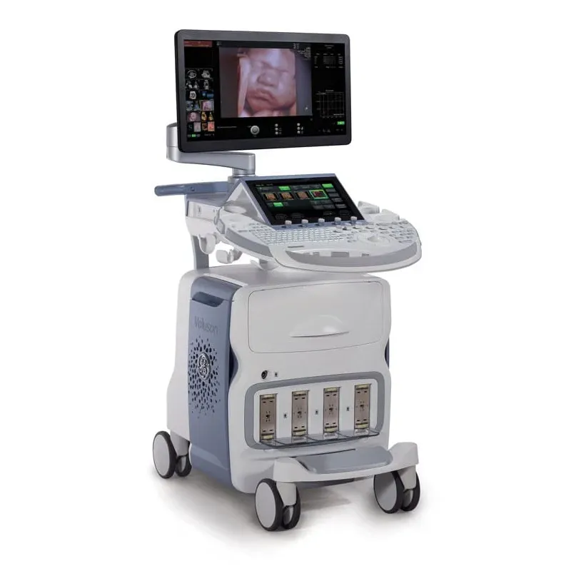
Voluson E10 (GE Healthcare): premium women's health ultrasound with Radiance System Architecture, HDlive/HDlive Studio 3D/4D rendering, Electronic 4D, SonoNT/SonoBiometry automation, Tricefy cloud sharing, wide range of matrix & HF probes.
The GE VOLUSON E10 is a premium ultrasound system designed for advanced OB/GYN imaging. Built with Radiance System Architecture and advanced HDlive Rendering Technology, it delivers exceptional 2D, 3D, and real-time 4D imaging performance for a wide range of women's health applications.
Designed to support everything from routine obstetric examinations to complex fetal and gynecological assessments, the VOLUSON E10 combines advanced processing power, high-resolution imaging, intuitive workflow tools, and sophisticated volume imaging technologies.
The GE VOLUSON E10 represents a high level of ultrasound imaging technology, offering advanced capabilities in 2D, 3D, and 4D imaging. Its powerful imaging platform is designed to provide excellent penetration, high-resolution visualization, and detailed anatomical information.
With increased data transfer capabilities and advanced processing technology, the system supports high image resolution and fast frame rates, helping clinicians obtain clear and detailed images efficiently.
At the core of the VOLUSON E10 is the Radiance System Architecture, designed to provide powerful processing capabilities and advanced imaging performance. This platform supports demanding clinical applications, from routine obstetric imaging to complex fetal echocardiography.
When combined with compatible advanced transducers, including the eM6C, the Radiance architecture supports ultra-fast volume rates, flexible imaging formats, and excellent image resolution for detailed volumetric imaging.
The VOLUSON E10 features advanced display technology designed to provide clear visualization and intuitive system control.
The VOLUSON E10 is designed to streamline the imaging workflow through intuitive navigation and efficient patient information management.
The electronic 4D imaging system provides real-time visualization of three-dimensional anatomical structures. This technology allows clinicians to assess complex anatomy and dynamic processes with excellent clarity and detail.
Bi-Plane Imaging allows simultaneous visualization of high-resolution images in two perpendicular planes. This capability provides complementary anatomical views and supports more comprehensive examination and evaluation.
Volume Contrast Imaging (VCI-A) is designed to enhance contrast resolution by providing improved clarity and detail throughout a thick-slice imaging volume. This technology can assist in the visualization of complex anatomy and subtle tissue differences.
eSTIC (Spatio-Temporal Image Correlation) is designed to support efficient fetal cardiac imaging by acquiring volumetric datasets that can be used for detailed evaluation of the fetal heart.
By streamlining the acquisition process, eSTIC helps improve workflow efficiency while maintaining detailed imaging information for advanced fetal cardiac assessment.
e4D SnapShot provides convenient access to high-resolution 3D volumes or eSTIC datasets directly from real-time 4D imaging. With simplified access to volumetric data, this feature helps streamline examinations while supporting comprehensive image acquisition.
V-SRI (Volume Speckle Reduction Imaging) is designed to improve 3D and 4D image quality by reducing speckle artifacts and improving the smoothness of rendered images.
Advanced VCI allows adjustment of slice thickness within 3D and 4D datasets to enhance contrast resolution and improve visualization, particularly when evaluating complex anatomical structures.
OmniView allows clinicians to generate customized imaging planes from a 3D or 4D volume dataset. By tracing a line, curve, polyline, or contour through a structure, users can obtain views that may be difficult to achieve with conventional 2D imaging.
SonoRenderlive simplifies volume rendering by automatically optimizing the placement of the rendering line for improved surface visualization. During real-time 4D examinations, the system can continuously adapt to movement, helping maintain optimal visualization of dynamic structures.
Combined with advanced rendering technologies such as HDlive and HDlive Studio, the VOLUSON E10 provides highly detailed volumetric visualization for advanced clinical evaluation.
The GE VOLUSON E10 combines advanced processing power, Radiance System Architecture, sophisticated 2D/3D/4D imaging, HDlive rendering, automated measurement tools, and comprehensive workflow solutions.
With technologies such as SonoNT, SonoBiometry, SonoVCAD, HDlive Studio, VCI-A, eSTIC, OmniView, SonoRenderlive, and ViewPoint Reporting, the system provides a comprehensive imaging platform for demanding obstetric and gynecological applications.
The GE VOLUSON E10 is a premium ultrasound system designed for advanced OB/GYN imaging. Built with Radiance System Architecture and advanced HDlive Rendering Technology, it delivers exceptional 2D, 3D, and real-time 4D imaging performance for a wide range of women's health applications.
Designed to support everything from routine obstetric examinations to complex fetal and gynecological assessments, the VOLUSON E10 combines advanced processing power, high-resolution imaging, intuitive workflow tools, and sophisticated volume imaging technologies.
The GE VOLUSON E10 represents a high level of ultrasound imaging technology, offering advanced capabilities in 2D, 3D, and 4D imaging. Its powerful imaging platform is designed to provide excellent penetration, high-resolution visualization, and detailed anatomical information.
With increased data transfer capabilities and advanced processing technology, the system supports high image resolution and fast frame rates, helping clinicians obtain clear and detailed images efficiently.
At the core of the VOLUSON E10 is the Radiance System Architecture, designed to provide powerful processing capabilities and advanced imaging performance. This platform supports demanding clinical applications, from routine obstetric imaging to complex fetal echocardiography.
When combined with compatible advanced transducers, including the eM6C, the Radiance architecture supports ultra-fast volume rates, flexible imaging formats, and excellent image resolution for detailed volumetric imaging.
The VOLUSON E10 features advanced display technology designed to provide clear visualization and intuitive system control.
The VOLUSON E10 is designed to streamline the imaging workflow through intuitive navigation and efficient patient information management.
The electronic 4D imaging system provides real-time visualization of three-dimensional anatomical structures. This technology allows clinicians to assess complex anatomy and dynamic processes with excellent clarity and detail.
Bi-Plane Imaging allows simultaneous visualization of high-resolution images in two perpendicular planes. This capability provides complementary anatomical views and supports more comprehensive examination and evaluation.
Volume Contrast Imaging (VCI-A) is designed to enhance contrast resolution by providing improved clarity and detail throughout a thick-slice imaging volume. This technology can assist in the visualization of complex anatomy and subtle tissue differences.
eSTIC (Spatio-Temporal Image Correlation) is designed to support efficient fetal cardiac imaging by acquiring volumetric datasets that can be used for detailed evaluation of the fetal heart.
By streamlining the acquisition process, eSTIC helps improve workflow efficiency while maintaining detailed imaging information for advanced fetal cardiac assessment.
e4D SnapShot provides convenient access to high-resolution 3D volumes or eSTIC datasets directly from real-time 4D imaging. With simplified access to volumetric data, this feature helps streamline examinations while supporting comprehensive image acquisition.
V-SRI (Volume Speckle Reduction Imaging) is designed to improve 3D and 4D image quality by reducing speckle artifacts and improving the smoothness of rendered images.
Advanced VCI allows adjustment of slice thickness within 3D and 4D datasets to enhance contrast resolution and improve visualization, particularly when evaluating complex anatomical structures.
OmniView allows clinicians to generate customized imaging planes from a 3D or 4D volume dataset. By tracing a line, curve, polyline, or contour through a structure, users can obtain views that may be difficult to achieve with conventional 2D imaging.
SonoRenderlive simplifies volume rendering by automatically optimizing the placement of the rendering line for improved surface visualization. During real-time 4D examinations, the system can continuously adapt to movement, helping maintain optimal visualization of dynamic structures.
Combined with advanced rendering technologies such as HDlive and HDlive Studio, the VOLUSON E10 provides highly detailed volumetric visualization for advanced clinical evaluation.
The GE VOLUSON E10 combines advanced processing power, Radiance System Architecture, sophisticated 2D/3D/4D imaging, HDlive rendering, automated measurement tools, and comprehensive workflow solutions.
With technologies such as SonoNT, SonoBiometry, SonoVCAD, HDlive Studio, VCI-A, eSTIC, OmniView, SonoRenderlive, and ViewPoint Reporting, the system provides a comprehensive imaging platform for demanding obstetric and gynecological applications.
 Francais