Phone: (+33) 625 16 20 71 - Sunday to Friday
contact@alliancemedicalfrance.fr
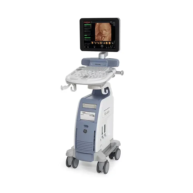
GE Voluson P8 is a women’s health ultrasound system offering 2D imaging, Color and Pulsed Doppler, 3D/4D imaging, workflow tools, and advanced technologies such as SRI, CrossXBeamCRI and HD-Flow
The GE Voluson P8 is an ultrasound system designed to support women's health imaging applications. Building on the established Voluson platform, it combines image quality, volumetric imaging capabilities, workflow tools, and an ergonomic design in a cost-effective solution.
As an entry point into the Voluson family, the system provides practical imaging capabilities that can be adapted to the needs of different clinical practices, patient populations, and budgets. Software upgrade options may also allow the system's capabilities to evolve according to changing clinical requirements.
The Voluson P8 features an ergonomic keyboard designed to improve user comfort and help reduce unnecessary movement during extended scanning sessions.
The Voluson P8 provides essential imaging modes for routine women's health examinations, including high-quality 2D imaging, Color Doppler, and Pulsed-Wave Doppler. Optional volumetric imaging capabilities provide additional anatomical information for advanced examinations.
Automatic optimization tools for 2D and Doppler imaging help improve image settings with minimal manual adjustment. Automatic focus positioning can also simplify the placement of the focal zone, allowing the operator to concentrate on the examination.
The Voluson P8 incorporates advanced imaging technologies designed to improve visualization and support diagnostic assessment.
The system supports 3D/4D imaging for the acquisition, visualization, and analysis of volumetric data. Volumes can be reviewed during the examination or analyzed offline using different sectional planes and rendering techniques.
The system supports a wide-sector field of view with compatible endocavitary probes, allowing more anatomical information to be displayed within a single image compared with a standard field of view.
Raw data storage allows acquired imaging information to be retained for further review and post-processing. This feature can support virtual rescanning and additional image adjustments after the examination.
The Voluson P8 includes reporting and archiving tools designed to simplify examination documentation and result management.
Combining 2D imaging, Doppler capabilities, advanced image optimization, volumetric imaging tools, reporting functions, and an ergonomic workflow, the GE Voluson P8 provides a versatile ultrasound solution for obstetric and gynecological imaging environments.
Please note: This offer includes the ultrasound system only. Probes and accessories are not included. Please contact us to discuss available probe options.
The GE Voluson P8 is an ultrasound system designed to support women's health imaging applications. Building on the established Voluson platform, it combines image quality, volumetric imaging capabilities, workflow tools, and an ergonomic design in a cost-effective solution.
As an entry point into the Voluson family, the system provides practical imaging capabilities that can be adapted to the needs of different clinical practices, patient populations, and budgets. Software upgrade options may also allow the system's capabilities to evolve according to changing clinical requirements.
The Voluson P8 features an ergonomic keyboard designed to improve user comfort and help reduce unnecessary movement during extended scanning sessions.
The Voluson P8 provides essential imaging modes for routine women's health examinations, including high-quality 2D imaging, Color Doppler, and Pulsed-Wave Doppler. Optional volumetric imaging capabilities provide additional anatomical information for advanced examinations.
Automatic optimization tools for 2D and Doppler imaging help improve image settings with minimal manual adjustment. Automatic focus positioning can also simplify the placement of the focal zone, allowing the operator to concentrate on the examination.
The Voluson P8 incorporates advanced imaging technologies designed to improve visualization and support diagnostic assessment.
The system supports 3D/4D imaging for the acquisition, visualization, and analysis of volumetric data. Volumes can be reviewed during the examination or analyzed offline using different sectional planes and rendering techniques.
The system supports a wide-sector field of view with compatible endocavitary probes, allowing more anatomical information to be displayed within a single image compared with a standard field of view.
Raw data storage allows acquired imaging information to be retained for further review and post-processing. This feature can support virtual rescanning and additional image adjustments after the examination.
The Voluson P8 includes reporting and archiving tools designed to simplify examination documentation and result management.
Combining 2D imaging, Doppler capabilities, advanced image optimization, volumetric imaging tools, reporting functions, and an ergonomic workflow, the GE Voluson P8 provides a versatile ultrasound solution for obstetric and gynecological imaging environments.
Please note: This offer includes the ultrasound system only. Probes and accessories are not included. Please contact us to discuss available probe options.
 Francais