Phone: (+33) 625 16 20 71 - Sunday to Friday contact@alliancemedicalfrance.fr
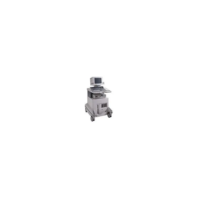
Philips Echographe HD 1500

Philips

Echographe HD 1500

Philips HDI 1500 is a versatile ultrasound system featuring 2D imaging, M-Mode, Color and Power Doppler, pulsed and continuous-wave Doppler, optional 3D imaging, DICOM connectivity, and image storage.

Philips HDI 1500 Ultrasound System

The HDI 1500 is a versatile ultrasound system designed for a wide range of clinical applications. It uses digital broadband technology and supports linear, convex, multi-beam, and static transducers.

Imaging Modes

  • 2D grayscale imaging.
  • M-Mode and Color M-Mode.
  • Color Doppler.
  • Power Angio® Color.
  • Pulsed-Wave Doppler.
  • Continuous-Wave Doppler.
  • Triple simultaneous imaging.
  • Optional 3D imaging.

Clinical Applications

  • Adult cardiology.
  • Obstetrics.
  • Gynecology.
  • Urology.
  • General radiology.

Storage and Connectivity

With integrated image compression, the HDI 1500 can store up to approximately 4,000 images, depending on file size. Images can be recalled and exported in JPEG or BMP formats.

  • DICOM connectivity.
  • Internal image and patient data storage.
  • Data transfer to removable media.
  • Compatible black-and-white and color printers.
  • Report generation and printing.

System Technology

The HDI 1500 is based on a Pentium computer platform combined with dedicated ultrasound electronics. It uses digital beamforming, echo processing, Color and Continuous-Wave Doppler processing, and digital scan conversion to generate ultrasound images.

  • Pentium-based computer platform.
  • Digital beamformer.
  • Echo processor.
  • Color Doppler processor.
  • Continuous-Wave Doppler processor.
  • Digital scan converter.
  • Real-time operating system.
Quick delivery worldwide
All our equipment is rigorously tested to ensure its quality and compliance with safety standards.
Download brochure

Philips HDI 1500 Ultrasound System

The HDI 1500 is a versatile ultrasound system designed for a wide range of clinical applications. It uses digital broadband technology and supports linear, convex, multi-beam, and static transducers.

Imaging Modes

  • 2D grayscale imaging.
  • M-Mode and Color M-Mode.
  • Color Doppler.
  • Power Angio® Color.
  • Pulsed-Wave Doppler.
  • Continuous-Wave Doppler.
  • Triple simultaneous imaging.
  • Optional 3D imaging.

Clinical Applications

  • Adult cardiology.
  • Obstetrics.
  • Gynecology.
  • Urology.
  • General radiology.

Storage and Connectivity

With integrated image compression, the HDI 1500 can store up to approximately 4,000 images, depending on file size. Images can be recalled and exported in JPEG or BMP formats.

  • DICOM connectivity.
  • Internal image and patient data storage.
  • Data transfer to removable media.
  • Compatible black-and-white and color printers.
  • Report generation and printing.

System Technology

The HDI 1500 is based on a Pentium computer platform combined with dedicated ultrasound electronics. It uses digital beamforming, echo processing, Color and Continuous-Wave Doppler processing, and digital scan conversion to generate ultrasound images.

  • Pentium-based computer platform.
  • Digital beamformer.
  • Echo processor.
  • Color Doppler processor.
  • Continuous-Wave Doppler processor.
  • Digital scan converter.
  • Real-time operating system.

Technology and Performance

  • Digital Ultrasound Technology: The HDI 1500 incorporates broadband digital ultrasound technology designed for a wide range of clinical applications.
  • Versatile Transducers: Compatible with various transducer configurations, including linear and convex probes, to meet different examination requirements.
  • 3D Imaging: The optional three-dimensional imaging capability provides detailed visualization of anatomical structures.
  • Storage and Export: The system allows image storage and retrieval, with the ability to export images to a computer in compatible image formats.
  • DICOM Connectivity: Facilitates integration into digital healthcare environments and the exchange of medical imaging information.
  • Printing Options: Supports compatible printing solutions for producing diagnostic images.

Linear Array Probes

  • L7-4 – Linear array probe.
  • L12-5 – Linear array probe.
  • CL10-5 – Linear array probe with synthetic aperture and trapezoidal imaging (version 2.01.00 and later).

Convex Array Probes

  • C5-2 – Convex array probe.
  • C7-4 – Convex array probe.
  • C8-4v – Convex array probe.
  • C8-5 – Convex array probe.
  • C9-5 – Convex array probe.

Phased Array Probes

  • P4-2 – Phased-array probe.
  • P7-4 – Phased-array probe.
  • P5-3 – Phased-array probe with Tissue Harmonic Imaging (version 2.01.00 and later).
Scroll