Phone: (+33) 625 16 20 71 - Sunday to Friday contact@alliancemedicalfrance.fr
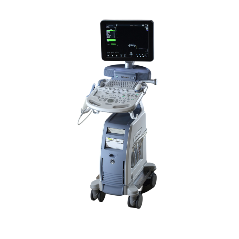
GE HealthCare Échographe VOLUSON P6

GE HealthCare

Échographe VOLUSON P6

GE Voluson P6: entry-level Voluson ultrasound for OB/GYN and fertility. Features 2D/3D/4D imaging, SRI, CrossXBeam, BetaView, HD-Zoom, wide 2D probe range, wireless connectivity, upgrade pathways.

Overview

The GE Voluson P6 is a high-performance ultrasound system designed primarily for obstetrics and gynecology. Its compact and cost-effective design makes it suitable for clinics and healthcare facilities with limited space and budget.

Imaging Quality

The system provides high-quality 2D imaging for routine obstetric and gynecological examinations. Technologies such as CrossXBeam and Speckle Reduction Imaging (SRI) enhance tissue visualization, improve image clarity, and reduce image noise.

3D/4D Imaging

The Voluson P6 supports real-time 3D and 4D imaging, making it suitable for fertility assessments, embryo evaluation, and embryo transfer procedures. Multiple rendering methods allow detailed volume analysis and visualization of anatomical structures.

Workflow and Automation

  • Intuitive user interface with simplified keyboard layout
  • 17-inch HD LCD display
  • Independent monitor adjustment for improved ergonomics
  • Automatic Optimization (AO) for rapid image adjustment
  • Three active probe ports

Compatible Probe Types

The system supports a range of probes, including convex, linear, and endocavitary probes. Probes are not included with this configuration.

Advanced Options

  • SonoNT™
  • SonoAVC™
  • SonoVCAD
  • Color Imaging
  • DICOM connectivity

Design and Mobility

Weighing approximately 65 kg, the Voluson P6 has a compact footprint that makes it easy to install and use in space-constrained clinical environments. Its ergonomic design provides convenient access to controls and connected probes.

Quick delivery worldwide
All our equipment is rigorously tested to ensure its quality and compliance with safety standards.
Download brochure

Overview

The GE Voluson P6 is a high-performance ultrasound system designed primarily for obstetrics and gynecology. Its compact and cost-effective design makes it suitable for clinics and healthcare facilities with limited space and budget.

Imaging Quality

The system provides high-quality 2D imaging for routine obstetric and gynecological examinations. Technologies such as CrossXBeam and Speckle Reduction Imaging (SRI) enhance tissue visualization, improve image clarity, and reduce image noise.

3D/4D Imaging

The Voluson P6 supports real-time 3D and 4D imaging, making it suitable for fertility assessments, embryo evaluation, and embryo transfer procedures. Multiple rendering methods allow detailed volume analysis and visualization of anatomical structures.

Workflow and Automation

  • Intuitive user interface with simplified keyboard layout
  • 17-inch HD LCD display
  • Independent monitor adjustment for improved ergonomics
  • Automatic Optimization (AO) for rapid image adjustment
  • Three active probe ports

Compatible Probe Types

The system supports a range of probes, including convex, linear, and endocavitary probes. Probes are not included with this configuration.

Advanced Options

  • SonoNT™
  • SonoAVC™
  • SonoVCAD
  • Color Imaging
  • DICOM connectivity

Design and Mobility

Weighing approximately 65 kg, the Voluson P6 has a compact footprint that makes it easy to install and use in space-constrained clinical environments. Its ergonomic design provides convenient access to controls and connected probes.

  • High-quality 2D imaging for routine gynecological and obstetric examinations
  • Real-time 3D/4D imaging for fertility, embryo, and embryo transfer assessments
  • CrossXBeam for improved tissue resolution and interface differentiation
  • Speckle Reduction Imaging (SRI) to reduce image noise and enhance clarity
  • HD-Zoom for easy localization of regions of interest and detailed examination
  • Automatic Optimization (AO) of imaging parameters for consistent image quality
  • Intuitive interface with a simplified keyboard for fast and easy navigation
  • High-resolution 17-inch LCD display
  • Three active probe ports for maximum flexibility
  • Compatible with convex, linear, and endocavitary probes
  • Optional software including SonoNT™, SonoAVC™, and SonoVCAD for advanced analysis
  • Compact and lightweight design suitable for space-constrained environments
  • Integrated DVR module for recording and streaming examinations
  • DICOM 3.0 connectivity for easy integration with medical systems
  • Integrated probe holder for probe protection and convenient positioning
  • Affordable software upgrades allowing the system to evolve with future needs
Encombrement minimal :
Design compact et léger pour une installation facile dans des espaces réduits
Options logicielles :
SonoNT™, SonoAVC™, SonoVCAD, STIC, Utérus VCI, IOTA, Omniview
Connectivité :
DICOM 3.0 pour une intégration facile avec d'autres systèmes
Fonctionnalités automatiques :
Optimisation automatique (AO) pour une meilleure qualité d'image
Technologies d'imagerie :
Imagerie 2D de haute qualitéImagerie 3D et 4D en temps réelCrossXBeam (amélioration de la résolution des tissus)Speckle Reduction Imaging (SRI, réduction du bruit de l'image)HD-Zoom (localisation des régions d'intérêt)
Type de sonde :
Sondes abdominales volumétriques RAB6-D 3D/4D de nouvelle génération
Sondes :
3 ports de sondes actifs, compatibles avec des sondes convexes, linéaires et endocavitaires
Écran :
LCD de 17 pouces, haute résolution
Poids :
65 kg
Dimensions :
49,5 cm x 67 cm x 42 cm
Alimentation :
Fonctionnement sur secteur
Protection des sondes :
Support de sonde pour une protection optimale et un positionnement facile
  • RAB2-6-RS — convex volume probe, used for 2D/3D fetal brain, abdomen, spine, and face imaging
  • 4C-RS — curved probe, used for 2D uterus imaging
  • RIC5-9W-RS — endocavitary volume probe, used for cyst imaging, wide sector, 3D uterus views, and Tomographic Ultrasound Imaging (TUI)
Scroll