Phone: (+33) 625 16 20 71 - Sunday to Friday
contact@alliancemedicalfrance.fr
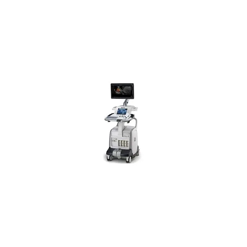
GE Vivid E9: 4D cardiovascular ultrasound with Accelerated Volume Architecture (8X processing power), single-beat full-volume capture, 4D quantification (Auto LVQ, Strain), 4D Stress, and an ergonomic, mobile design for bedside cardiac imaging.
Box only, price without probes. Contact us for probe options.
The GE Healthcare Vivid™ E9 is a groundbreaking cardiovascular ultrasound system designed to deliver exceptional-quality 4D images. This device is specifically developed for practitioners seeking an ergonomic solution for cardiac diagnosis.
Thanks to its Accelerated Volume Architecture (AVA) platform, the Vivid™ E9 offers 8 times the processing power of traditional ultrasound systems, enabling the acquisition of complete data volumes in one or several heartbeats. This system reduces artifacts while eliminating the need for ECG synchronization, making 4D imaging more accessible and fluid.
One of the key strengths of the Vivid™ E9 is its ability to capture 4D volumes with exceptional precision and speed, providing detailed views of the entire heart, including areas that are difficult to reach with traditional ultrasound systems.
Ease of use is a priority with the Vivid™ E9, whose lightweight and compact console is designed to be easily moved for use in an office setting or at the patient's bedside.
The integration of Scan Assist and Scan Assist Pro enables simplified management of examination protocols, whether for traditional, cardiac, vascular, or abdominal ultrasounds. These tools reduce the number of manipulations required, ensuring better reproducibility and consistent image acquisition.
4D quantification on the Vivid™ E9 includes powerful tools for accurate and reproducible diagnosis.
Box only, price without probes. Contact us for probe options.
The GE Healthcare Vivid™ E9 is a groundbreaking cardiovascular ultrasound system designed to deliver exceptional-quality 4D images. This device is specifically developed for practitioners seeking an ergonomic solution for cardiac diagnosis.
Thanks to its Accelerated Volume Architecture (AVA) platform, the Vivid™ E9 offers 8 times the processing power of traditional ultrasound systems, enabling the acquisition of complete data volumes in one or several heartbeats. This system reduces artifacts while eliminating the need for ECG synchronization, making 4D imaging more accessible and fluid.
One of the key strengths of the Vivid™ E9 is its ability to capture 4D volumes with exceptional precision and speed, providing detailed views of the entire heart, including areas that are difficult to reach with traditional ultrasound systems.
Ease of use is a priority with the Vivid™ E9, whose lightweight and compact console is designed to be easily moved for use in an office setting or at the patient's bedside.
The integration of Scan Assist and Scan Assist Pro enables simplified management of examination protocols, whether for traditional, cardiac, vascular, or abdominal ultrasounds. These tools reduce the number of manipulations required, ensuring better reproducibility and consistent image acquisition.
4D quantification on the Vivid™ E9 includes powerful tools for accurate and reproducible diagnosis.
Advanced 4D Imaging: Delivers full 4D volumes with 8 times greater processing power thanks to Accelerated Volume Architecture (AVA) technology.
Exceptional Image Quality: Precise resolution and high-quality definition, enabling detailed assessment of the heart, valves, and endocardial walls.
Optimized Ergonomics: Lightweight, compact console that's easy to move between rooms, with an adjustable floating keyboard for personalized comfort.
Intuitive Interface: 17-inch LCD touchscreen and a secondary 10-inch display, offering smooth navigation and instant image visualization.
Workflow Automation Tools: Integration of Scan Assist and Scan Assist Pro to simplify image acquisition and the application of customized protocols.
High-Performance Probes: Compatible with the latest generation GE Healthcare probes, such as the M5Sc-D probe, delivering exceptionally clear images.
Integrated 2D and 4D Imaging: Combines the benefits of classic 2D imaging with the power of 4D imaging for a complete and precise analysis of the heart.
Quantification: 4D quantification tools, such as 4D Auto LVQ, for precise analysis of volumes, ejection fractions, and ventricular masses.
Real-Time View: Instant 4D visualization with tools like 4D Views, allowing precise views to be selected and displayed directly without complex manual manipulation.
Portability and Mobility: Compact, rugged design, ideal for bedside use while still delivering optimal image quality.
Optimized Stress Exams: 4D Stress function that allows full volumes to be acquired under stress and analyzed from different perspectives, for a thorough evaluation.
Simplified Data Management: Streamlined data storage with multiple USB ports and a DVR recorder, for smooth management of patient studies.
Adaptability: Support for multiple protocols for cardiac, vascular, and abdominal exams, with a customizable interface tailored to clinician needs.
4V-D probe: Second-generation 4D probe dedicated to full cardiac volume acquisition.
M5S probe: Matrix probe combining matrix-array and single-crystal technologies for exceptional endocardial definition and image texture.
D-series transducers: New generation of probes enhancing 2D, 4D and shared-service imaging across your entire patient population.
12S probe: Probe used for color Doppler imaging (pulmonary artery, carotid) and vascular exams.
Multiple USB ports: For fast, simplified patient data management.
DVR recorder: Built in for archiving studies and exam videos.
 Francais