Phone: (+33) 625 16 20 71 - Sunday to Friday contact@alliancemedicalfrance.fr
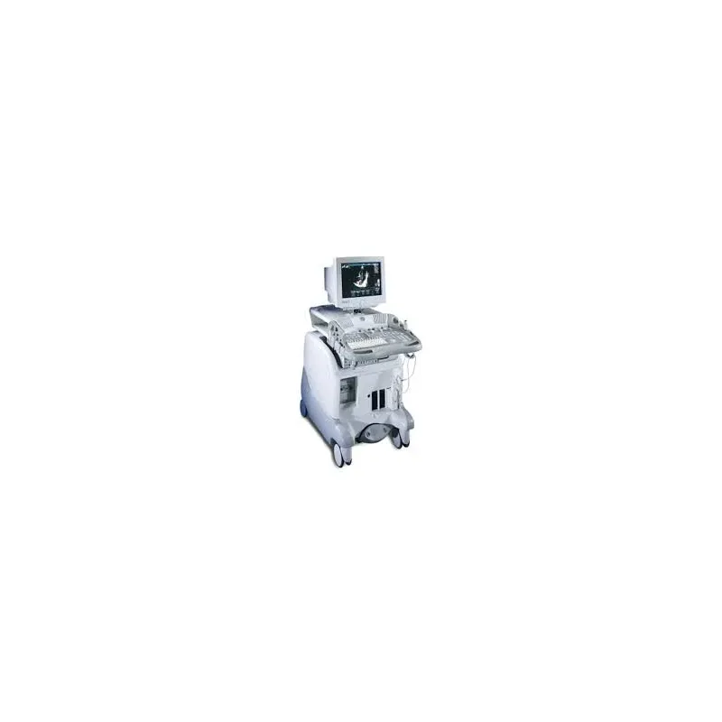
GE HealthCare Système d'échographie cardiovasculaire Vivid 3

GE HealthCare

Système d'échographie cardiovasculaire Vivid 3

Starting from
3 890 .00 €

GE Vivid 3 is a versatile cardiovascular ultrasound system offering high-quality imaging, advanced 3D, Doppler, raw-data processing, and broad clinical applications.

GE Vivid 3

The GE Vivid 3 is a high-performance ultrasound system primarily designed for cardiovascular applications. It delivers excellent image quality, flexible operation, and advanced features for fast and reliable examinations.

Imaging Technology

The Vivid 3 incorporates TruScan technology, which captures and stores raw image data. This allows users to review, measure, optimize, and analyze images later while preserving their original quality.

The system also provides high frame rates and enhanced color-flow sensitivity, making it particularly suitable for cardiac and vascular examinations.

Automatic Optimization

ATO (Automatic Tissue Optimization) automatically adjusts imaging parameters to quickly achieve optimal image quality. This feature simplifies examinations and helps improve workflow efficiency.

Clinical Applications

  • Cardiology and cardiovascular imaging
  • Vascular imaging
  • Gynecology
  • Pediatrics
  • Abdominal imaging
  • Intraoperative applications

Advanced Features

  • Stress Echo for cardiovascular stress examinations
  • DICOM for medical data exchange and archiving
  • MPEGvue for recording and transferring patient files
  • Support for a wide range of probes and transducers

Connectivity and Data Management

With DICOM connectivity, the GE Vivid 3 facilitates the transfer, archiving, and management of patient data within healthcare information systems.

Quick delivery worldwide
All our equipment is rigorously tested to ensure its quality and compliance with safety standards.
Download brochure

GE Vivid 3

The GE Vivid 3 is a high-performance ultrasound system primarily designed for cardiovascular applications. It delivers excellent image quality, flexible operation, and advanced features for fast and reliable examinations.

Imaging Technology

The Vivid 3 incorporates TruScan technology, which captures and stores raw image data. This allows users to review, measure, optimize, and analyze images later while preserving their original quality.

The system also provides high frame rates and enhanced color-flow sensitivity, making it particularly suitable for cardiac and vascular examinations.

Automatic Optimization

ATO (Automatic Tissue Optimization) automatically adjusts imaging parameters to quickly achieve optimal image quality. This feature simplifies examinations and helps improve workflow efficiency.

Clinical Applications

  • Cardiology and cardiovascular imaging
  • Vascular imaging
  • Gynecology
  • Pediatrics
  • Abdominal imaging
  • Intraoperative applications

Advanced Features

  • Stress Echo for cardiovascular stress examinations
  • DICOM for medical data exchange and archiving
  • MPEGvue for recording and transferring patient files
  • Support for a wide range of probes and transducers

Connectivity and Data Management

With DICOM connectivity, the GE Vivid 3 facilitates the transfer, archiving, and management of patient data within healthcare information systems.

  • TruScan Technology for raw image acquisition, ensuring optimal image quality and integrity.
  • Automatic Tissue Optimization (ATO) for obtaining high-quality images quickly and easily.
  • Wide range of compatible probes for cardiac, vascular, pediatric, and obstetric/gynecological applications.
  • DICOM connectivity for fast data transfer and efficient patient record management.
  • Stress echocardiography for rapid examinations with protocol changes during the examination.
  • MPEGvue recording for saving patient records, including high-resolution images, to CD.
  • Intuitive and simplified user interface for easy operation, even for clinicians who are not ultrasound specialists.
  • Advanced imaging options including coded harmonics, color Doppler, and PW/CW spectral Doppler for accurate blood-flow analysis.
  • B/M and M modes for dynamic analysis of the heart and soft tissues.
  • Customizable calculations and reports for cardiovascular and vascular applications, supporting accurate documentation.
  • Flexibility with up to 18 probes for a wide range of examination types.
  • 3D options for vascular studies, helping improve diagnostic accuracy.
  • Lightweight and compact system for easy installation and mobility across different clinical environments.
  • High performance with fast examination speeds and high image resolution for maximum productivity.
  • Upgradeable design allowing new features to be added and the system to adapt to the future needs of the healthcare facility.
Options supplémentaires :
Echo Stress pour des examens plus rapides, option 3D pour les études vasculaires
Enregistrement :
Option MPEGvue pour l'enregistrement des dossiers patients sur CD
Gestion des données :
TruAccess pour la gestion complète des données et stockage des images
Mode de fonctionnement :
B, B/M, M Mode
Calculs et rapports :
Logiciels pour calculs cardiovasculaires et vasculaires, rapports personnalisables
Options d'image :
Harmoniques codées, Doppler couleur, B-color, Doppler spectral PW/CW
Optimisation automatique :
Fonction d'optimisation automatique des tissus (ATO) pour des images de haute qualité
Technologie d'imagerie :
TruScan pour l'acquisition d'images brutes et l'optimisation des données
Connectivité :
DICOM pour l'exportation des images et des données
Moniteur :
Écran CRT couleur de 15 pouces
Probes compatibles :
Jusqu'à 18 sondes différentes, incluant des sondes sectorielles, linéaires et CW non-imagées
Dimensions :
132 cm (H) x 64 cm (L) x 112 cm (P)
Poids :
159 kg
Application :
Cardiaque, vasculaire, pédiatrique, obstétrique/gynécologique, abdominal, et surveillance intra-opératoire
Compatibilité :
Compatible avec les sondes GE 3S, 5S, 7L, 10L, P2P CW et P6D CW non-imagées
  • 3S – Phased-array cardiac transducer
  • 5S – Phased-array cardiac and pediatric transducer
  • 7S – Phased-array cardiac transducer
  • 10S – High-frequency neonatal transducer
  • C358 – Curved-array transducer
  • C721 – Curved-array pediatric and neonatal transducer
  • 7L – Linear transducer for vascular and superficial applications
  • 10L – Linear transducer for vascular and superficial imaging
  • 12L – Linear transducer for vascular and small-parts imaging
  • 6T – Adult transesophageal echocardiography (TEE) transducer
  • 8T – Pediatric TEE transducer
  • 9T – Pediatric TEE transducer
  • E721 – Endovaginal transducer for OB/GYN applications
  • P2D – Continuous-wave Doppler pencil transducer
  • P6D – Continuous-wave Doppler pencil transducer
Scroll