Phone: (+33) 625 16 20 71 - Sunday to Friday contact@alliancemedicalfrance.fr
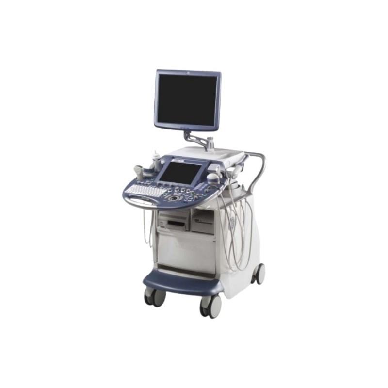
GE HealthCare Echographe Voluson E8 Expert

GE HealthCare

Echographe Voluson E8 Expert

GE Voluson E8/E8 Expert ultrasound system with 3D/4D volume scanning. Supports 2D, M-mode, spectral and color Doppler, B-Flow, XTD-View. Wide probe range, DICOM networking, digital archiving.

Overview

The Voluson E8 Expert is a premium ultrasound system designed for healthcare professionals who require high-quality imaging and advanced diagnostic capabilities. It supports a broad range of clinical applications, including obstetrics, gynecology, vascular, pediatric, musculoskeletal, abdominal, and neurological imaging.

Advanced Imaging

The system provides high-quality real-time imaging with advanced 3D and 4D capabilities. Technologies such as HDlive provide detailed, photo-realistic visualization, while VCI OmniView enables multi-angle volumetric analysis.

  • SonoAVC™ for automated follicle counting
  • STIC for advanced fetal cardiac assessment
  • VOCAL II for complex volume calculations
  • Elastography for tissue assessment
  • B-Flow for enhanced blood-flow visualization

Clinical Applications

  • Obstetrics and gynecology
  • Vascular imaging
  • Pediatric examinations
  • Musculoskeletal imaging
  • Abdominal examinations
  • Neurological imaging

Ergonomic Design

The Voluson E8 Expert features an ergonomic user interface with a 10.4-inch LCD touchscreen and a 19-inch high-resolution SXGA main display. Its adjustable floating keyboard can be positioned in three dimensions to improve comfort during extended examinations.

Connectivity and Data Management

The system supports up to four probe ports, with three active ports, and provides convenient connectivity for various peripherals. Internal image archiving with lossy or lossless compression is supported, along with data export through DVD, USB, or network connections.

Advanced post-processing allows 2D, 3D, and 4D images to be reviewed and adjusted while maintaining image quality. DICOM and IHE compatibility enables integration into modern hospital networks.

Quick delivery worldwide
All our equipment is rigorously tested to ensure its quality and compliance with safety standards.
Download brochure

Overview

The Voluson E8 Expert is a premium ultrasound system designed for healthcare professionals who require high-quality imaging and advanced diagnostic capabilities. It supports a broad range of clinical applications, including obstetrics, gynecology, vascular, pediatric, musculoskeletal, abdominal, and neurological imaging.

Advanced Imaging

The system provides high-quality real-time imaging with advanced 3D and 4D capabilities. Technologies such as HDlive provide detailed, photo-realistic visualization, while VCI OmniView enables multi-angle volumetric analysis.

  • SonoAVC™ for automated follicle counting
  • STIC for advanced fetal cardiac assessment
  • VOCAL II for complex volume calculations
  • Elastography for tissue assessment
  • B-Flow for enhanced blood-flow visualization

Clinical Applications

  • Obstetrics and gynecology
  • Vascular imaging
  • Pediatric examinations
  • Musculoskeletal imaging
  • Abdominal examinations
  • Neurological imaging

Ergonomic Design

The Voluson E8 Expert features an ergonomic user interface with a 10.4-inch LCD touchscreen and a 19-inch high-resolution SXGA main display. Its adjustable floating keyboard can be positioned in three dimensions to improve comfort during extended examinations.

Connectivity and Data Management

The system supports up to four probe ports, with three active ports, and provides convenient connectivity for various peripherals. Internal image archiving with lossy or lossless compression is supported, along with data export through DVD, USB, or network connections.

Advanced post-processing allows 2D, 3D, and 4D images to be reviewed and adjusted while maintaining image quality. DICOM and IHE compatibility enables integration into modern hospital networks.

Operating Modes

  • B, M, PW, CFM, PD, HD-Flow, TD, B-Flow
  • 3D/4D – Real-time 3D/4D imaging with SonoRenderStart

Imaging Technologies

  • HDlive
  • VCI OmniView
  • VOCAL II
  • SonoAVC™
  • SonoVCAD™
  • STIC

Post-Processing and Optimization

  • HD Zoom up to 22x
  • Gain adjustment, customizable grayscale maps and tint settings
  • Automatic Tissue Optimization (ATO)
  • Coded Harmonic Imaging
  • Compound Resolution Imaging (CRI)
  • Speckle Reduction with SRI II and V-SRI

User Interface and Patient Management

  • Multilingual user interface, including French
  • Integrated patient database
  • Automated measurement tools
  • Annotation with up to 400 storable terms
  • 117 body silhouettes for anatomical localization

Image and Data Management

  • 2D/3D Cine Loop with review and annotation functions
  • Export formats: DICOM, JPEG, AVI, BMP, MOV, and TIFF
  • Image compression: lossless or lossy according to preference
Protocoles supportés :
DICOM, DHCP, NetBIOS, Insite ExC
Interface réseau :
Ethernet, WLAN en option
Température d'utilisation :
18–30°C
Bruit émis :
< 55 dB
Ports de sonde :
4 (dont 3 actifs)
Disque dur intégré :
500 Go
Puissance max :
1000 VA
Alimentation :
100-240V AC / 50-60Hz
Poids :
env. 131 kg
Dimensions :
580 mm (L) x 920 mm (P) x 1393-1583 mm (H)
Écran secondaire :
Écran tactile LCD 10,4”
Écran principal :
LCD 19" SXGA (1280 x 1024), haute luminosité 220 cd/m²
Stockage :
USB, DVD+R(W)/CD-R(W), réseau
Profondeur d’exploration :
jusqu’à 36 cm
Dynamic Range :
274 dB
Colorimétrie :
24 bits, 16,8 millions de couleurs

2D Probes

  • 11L-D
  • 3S-D
  • 3Sp-D
  • 4C-D
  • 9L-D
  • AB2-7-D

3D/4D Volume Probes

  • RAB2-5-D
  • RAB4-8-D
  • RAB6-D
  • RAM3-8
  • RIC5-9-D
  • RIC6-12-D
Scroll