Phone: (+33) 625 16 20 71 - Sunday to Friday contact@alliancemedicalfrance.fr
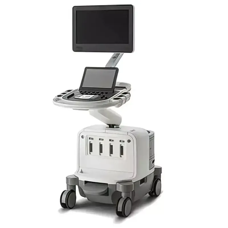
Philips Échographe EPIQ 7C

Philips

Échographe EPIQ 7C

The Philips EPIQ 7C is a premium cardiovascular ultrasound system with nSIGHT Imaging, xMATRIX transducers, Anatomical Intelligence (AIUS), HeartModel, Live 3D TEE, and EchoNavigator for interventional guidance.

Philips EPIQ 7C

The Philips EPIQ 7C is a premium cardiovascular ultrasound system designed to deliver excellent image quality, advanced quantification tools, and an efficient workflow for demanding clinical environments.

nSIGHT Imaging Technology

The proprietary nSIGHT Imaging architecture uses advanced digital acoustic beam reconstruction and optimized focusing to provide excellent pixel-level resolution and image uniformity.

  • Up to 30% increased penetration.
  • Up to 15% improved axial resolution.
  • Up to 4× higher volume rates with xMATRIX transducers.
  • 3D volume acquisition in a single heartbeat.
  • Reduced stitching artifacts during 3D imaging.

Advanced Imaging

The EPIQ 7C incorporates xMATRIX and PureWave transducer technologies, allowing smooth transitions between multiple imaging modes.

  • 2D imaging.
  • Doppler imaging.
  • M-mode.
  • Live xPlane.
  • Live 3D.
  • 3D Zoom.

Artificial Intelligence and Automated Quantification

Anatomical Intelligence (AIUS) enables automatic recognition of anatomical structures and facilitates reproducible measurements.

  • HeartModelA.I.: Automated quantification of cardiac volumes and function.
  • a2DQA.I.: Automated 2D cardiac quantitative analysis.
  • aCMQA.I.: Automated cardiac function analysis.
  • ZeroClick: Reduces manual interaction during examinations.
  • Mitral Valve NavigatorA.I.: Guided analysis and automated measurements of the mitral valve.

Interventional Imaging

EchoNavigator provides real-time synchronization between ultrasound and fluoroscopic images when integrated with Philips Allura Xper systems, supporting image-guided cardiovascular procedures.

Workflow and Ergonomics

  • Intuitive touchscreen interface.
  • Up to 80% fewer gestures required for operation.
  • Up to 15% fewer steps required for certain examinations.
  • SmartExam: Can reduce examination time by 30–50%.
  • Auto Doppler: Simplifies the vascular imaging workflow.
  • Articulated control panel and monitor for ergonomic positioning.
  • Cable hooks and recovery tray for improved organization.

Display and Mobility

  • MaxVue HD display: Provides an expanded viewing area for enhanced visualization.
  • Weight: Approximately 104 kg.
  • Designed for convenient movement within clinical environments.
  • Operating noise level of approximately 37–41 dB for a comfortable working environment.
Quick delivery worldwide
All our equipment is rigorously tested to ensure its quality and compliance with safety standards.
Download brochure

Philips EPIQ 7C

The Philips EPIQ 7C is a premium cardiovascular ultrasound system designed to deliver excellent image quality, advanced quantification tools, and an efficient workflow for demanding clinical environments.

nSIGHT Imaging Technology

The proprietary nSIGHT Imaging architecture uses advanced digital acoustic beam reconstruction and optimized focusing to provide excellent pixel-level resolution and image uniformity.

  • Up to 30% increased penetration.
  • Up to 15% improved axial resolution.
  • Up to 4× higher volume rates with xMATRIX transducers.
  • 3D volume acquisition in a single heartbeat.
  • Reduced stitching artifacts during 3D imaging.

Advanced Imaging

The EPIQ 7C incorporates xMATRIX and PureWave transducer technologies, allowing smooth transitions between multiple imaging modes.

  • 2D imaging.
  • Doppler imaging.
  • M-mode.
  • Live xPlane.
  • Live 3D.
  • 3D Zoom.

Artificial Intelligence and Automated Quantification

Anatomical Intelligence (AIUS) enables automatic recognition of anatomical structures and facilitates reproducible measurements.

  • HeartModelA.I.: Automated quantification of cardiac volumes and function.
  • a2DQA.I.: Automated 2D cardiac quantitative analysis.
  • aCMQA.I.: Automated cardiac function analysis.
  • ZeroClick: Reduces manual interaction during examinations.
  • Mitral Valve NavigatorA.I.: Guided analysis and automated measurements of the mitral valve.

Interventional Imaging

EchoNavigator provides real-time synchronization between ultrasound and fluoroscopic images when integrated with Philips Allura Xper systems, supporting image-guided cardiovascular procedures.

Workflow and Ergonomics

  • Intuitive touchscreen interface.
  • Up to 80% fewer gestures required for operation.
  • Up to 15% fewer steps required for certain examinations.
  • SmartExam: Can reduce examination time by 30–50%.
  • Auto Doppler: Simplifies the vascular imaging workflow.
  • Articulated control panel and monitor for ergonomic positioning.
  • Cable hooks and recovery tray for improved organization.

Display and Mobility

  • MaxVue HD display: Provides an expanded viewing area for enhanced visualization.
  • Weight: Approximately 104 kg.
  • Designed for convenient movement within clinical environments.
  • Operating noise level of approximately 37–41 dB for a comfortable working environment.
  • Live 3D Imaging: Advanced 3D imaging with xMATRIX transducers.
  • PureWave Technology: Enhanced image resolution and quality.
  • Anatomical Intelligence (AIUS): Integrated intelligent anatomical recognition.
  • Automated Quantification: HeartModelA.I., a2DQA.I., and aCMQA.I. applications.
  • Mitral Valve NavigatorA.I.: Advanced analysis of the mitral valve.
  • EchoNavigator Integration: Synchronization with fluoroscopic imaging.
  • Intuitive Ergonomic Interface: Designed for comfortable and efficient operation.
  • Streamlined Workflow: Reduced gestures and examination steps.
  • Quiet Operation: Suitable for demanding clinical environments.
  • SmartExam & Auto Doppler: Tools designed to improve clinical efficiency.
  • Proactive Monitoring: Supports remote maintenance and system monitoring.
Niveau sonore :
37–41 dB
Poids :
104 kg

xMATRIX / Live 3D transducers

  • X5-1 – used for one-beat Live 3D volumes
  • X8-2t – Live 3D transesophageal (TEE) transducer

Adult / pediatric echo

  • S5-1 – used for color compare and CW Doppler (VSD imaging)
  • S8-3 – pediatric probe (2D, color, AP4 views)

Vascular

  • L12-3 – linear probe, used for carotid/ICA PW Doppler
Scroll